MediVis project at Ostrava – CT scan

At this time I would like to introduce you to my project at IT4I National Supercomputing Center at Ostrava, Check. During this summer I was involved in a very interesting Post-processing and 3D visualization for medical 3D Visualization from CT images.
The Computed Tomography (CT) is a widespread technique used in medicine.With the help of image processing, visualization, and High Performance Computing (HPC) systems, we can further exploit the current CT technology to support a more precise and faster medical treatment. By achieving these tasks, we can reduce the very important aspect, which is the related risk of the treatment.
The project focuses on the segmentation, reconstruction, and 3D visualization of human organs from CT images. There is a cooperation between IT4I and medical doctors to help the evaluation of the state of the patients and support the treatment by the development of suitable techniques for 3D model reconstruction of selected human body organs. The CT scanning or so-called X-ray computed tomography (X-ray CT) could be considered as a non-invasive procedure since it allows us to see inside the human body without clinical surgery. The only limitation of the technology is the radiation dose during the process and its effects on the human body. The risk that comes from the radiation is much lower nowadays thanks to new types of CT machines.
Although CT technology is not new, the Italian radiologist Alessandro Vallebona was the first person who proposed a method in 1900s to represent a single slice of a body on a radiography film. This method was the so-called tomography which is well-known and widely investigated. The mathematical theory of the computed tomographic reconstruction can be traced to the invention of Radon transform (Johann Radon, 1917). He demonstrated mathematically, that from an infinite set of projections enough information to reconstruct a function.
This lead to CT and the first commercial scanners. The idea came from Sir Godfrey Hounsfield in 1967 , who invented the first CT and his solution was applied at the first EMI-Scanner was installed and used for brain scan from 1972 in Atkinson Morley Hospital in Wimbledon, England.
Of course, they were just the first ones, and we can continue this list including the name of the researchers and their results. The current CT scanners are largely upgraded, and the usage and investigation of the technology involve the math, physics, and information technology also.
The usage of CT scanners have been dramatically increased in the last few decades thanks to better resolution, the less harmful effect. Moreover nowadays there are different type of (tomography) machines for instance PET. You can see some pictures below that show, the CT machine, the slices, and a 3D liver based on CT results.
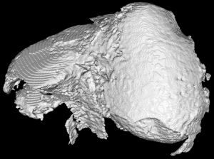
3D liver
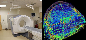
CT scanner and created slices

Leave a Reply