Medical image segmentation and visualization
Project reference: 1514
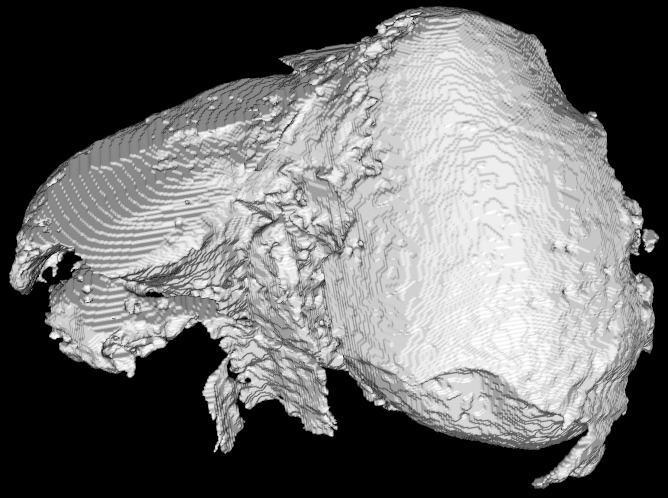
Project will focus on rapid prototyping and testing of different algorithms for image segmentation as well as visualization of 3D data. In a first stage implementation and testing of suitable image segmentation techniques for medical image segmentation will be done. As an input data for image segmentation the consecutive series of CT or MRI medical images will be used. After the segmentation visualization of the obtained 3D data will be performed. In the visualization stage focus will be on suitable pre-processing of the data for further 3D visualization
For rapid prototyping Matlab software will be used. Standard existing functions for image segmentation will be used. However there is possibility that new special functions will be developed and optimized using Matlab’s Parallel Computing Toolbox.
Project mentor: Petr Strakoš
Site Co-ordinator: Karina Pesatova
Learning Outcomes
Student will gain experience in:
- Image segmentation techniques
- 3D visualisation
- Matlab
- C, C++ programming
- using visualization software
Student Prerequisites (compulsory)
Programming skills in C, C++ and Matlab.
Student Prerequisites (desirable)
Image processing and segmentation, 3D data visualization, parallel processing.
Training Materials
Matlab webinars: http://www.mathworks.com/company/events/webinars/index.html?recordedwebinars
Segmentation & registration toolkit: http://www.itk.org
Visualization toolkit: http://www.vtk.org
Open source image segmentation software: http://www.itksnap.org/pmwiki/pmwiki.php, http://www.slicer.org/pages/Introduction, http://www.osirix-viewer.com/AboutOsiriX.html
Medical image databases: http://www.osirix-viewer.com/datasets/
Image segmentation algorithms: http://en.wikipedia.org/wiki/Region_growing, http://en.wikipedia.org/wiki/K-means_clustering, http://en.wikipedia.org/wiki/Flood_fill, http://en.wikipedia.org/wiki/Otsu%27s_method
Workplan
- Week 1: Training
- Week 2 : Work plan setting
- Week 3 – 6: Implementation and testing of image segmentation algorithms in Matlab
- Week 7 : Visualization of the results
- Week 8 : Final report completion and final presentation preparation
Final Product Description
Resulting visualizations and animations of the 3D models of internal organs can be used to demonstrate HPC capabilities to the public.
Adapting the Project – Increasing the Difficulty
By implementing more difficult image segmentation techniques and by developing the GUI.
Adapting the Project – Decreasing the Difficulty
By using already implemented functions in the image segmentation stage.
Resources
Software: Matlab, visualisation utilities (ParaView, Visit, Ensight, etc.), C, C++ programming environment
Hardware: high memory system, visualization server, anselm cluster
Access to the appropriate software and hardware will be provided by the IT4Innovations National Supercomputing Center.
Organization
